Femur ORIF Physical Therapy Protocol
This protocol guides rehabilitation post-femur fracture fixation, typically starting the day of surgery with prescribed physical therapy.
It addresses weight-bearing, range of motion, and strengthening,
progressing through phases to restore function and minimize complications.
Early intervention focuses on edema control, pain management, and gentle muscle activation,
while later stages emphasize advanced strengthening, proprioception, and a gradual return to activities.
Regular follow-up with the orthopedic surgeon is crucial, alongside adherence to the prescribed exercise program,
ensuring optimal recovery and long-term joint health.
This comprehensive physical therapy protocol outlines the rehabilitation process following open reduction and internal fixation (ORIF) of a femur fracture. It’s designed to provide a structured approach for clinicians and patients, aiming for optimal functional recovery and a safe return to activity. The protocol acknowledges the significant impact femoral fractures, particularly in the elderly, have on patient mobility and quality of life, necessitating a tailored rehabilitation program.
Post-operative care begins immediately, often with physical therapy initiated the same day as surgery, as per the surgeon’s prescription. This early intervention focuses on managing pain and swelling, initiating gentle range of motion exercises, and activating key muscle groups. The success of this protocol relies on a collaborative effort between the orthopedic surgeon, physical therapist, and the patient’s active participation.
This document serves as a guideline, and individual progression will vary based on fracture complexity, patient factors, and clinical judgment. Regular monitoring and adjustments to the program are essential to ensure optimal outcomes and prevent potential complications. The ultimate goal is to restore the patient’s pre-injury level of function, or as close as possible, while minimizing long-term disability.
Pre-Operative Considerations
Prior to femur ORIF surgery, optimizing the patient’s overall health is paramount for a successful rehabilitation. Addressing pre-existing conditions like cardiovascular disease, diabetes, or respiratory issues is crucial. Nutritional status should be assessed and optimized to support wound healing and recovery. Pre-operative physical therapy, if time allows, can focus on strengthening unaffected limbs and educating the patient about post-operative expectations.
A thorough understanding of the patient’s pre-injury functional level is essential to establish realistic goals. Identifying any pre-existing limitations or comorbidities will help tailor the rehabilitation program. Discussing potential weight-bearing restrictions and the need for assistive devices pre-operatively prepares the patient mentally and practically.
Patient education regarding the surgical procedure, potential complications, and the importance of adherence to the rehabilitation protocol is vital. Establishing a strong patient-therapist relationship before surgery fosters trust and encourages active participation in the recovery process. A home safety assessment may also be beneficial to minimize fall risk post-operatively.
Phase 1: Immediate Post-Operative (0-2 Weeks)
Initial focus is on pain and edema control, knee immobilization (often locked in extension), and gentle muscle activation exercises.
Weight-bearing is restricted as directed by the surgeon.
Weight-Bearing Restrictions
Weight-bearing status post-femur ORIF is dictated by fracture stability and surgeon preference. Initially, patients typically follow non-weight-bearing (NWB) or toe-touch weight-bearing (TTWB) for the first 0-6 weeks. This restriction protects the healing fracture site and allows for callus formation.
Strict adherence to weight-bearing precautions is paramount to avoid complications like non-union or hardware failure. Assistive devices, such as crutches or a walker, are essential during this phase. Progressive weight-bearing is introduced gradually, often starting with partial weight-bearing (PWB) at 25-50% of body weight, as tolerated and guided by pain levels and radiographic evidence of healing.
Regular monitoring by the physical therapist and surgeon is crucial to ensure appropriate progression. Full weight-bearing (FWB) is typically permitted around 6-12 weeks, contingent upon adequate fracture healing and sufficient muscle strength. Any increase in pain or instability warrants immediate evaluation.
Knee Immobilization
Post-operative knee immobilization following femur ORIF aims to protect the surgical site and minimize pain. A knee immobilizer, often locked in full extension, is commonly utilized for the initial 0-2 weeks. This restricts excessive movement and provides stability during the early healing stages.
The duration of immobilization varies based on fracture complexity and surgical technique. Prolonged immobilization can lead to muscle atrophy and joint stiffness, therefore, early controlled range of motion exercises are incorporated as tolerated. The immobilizer may be discontinued earlier if the fixation is stable and pain allows.
Patients are instructed on proper donning and doffing of the immobilizer and advised to avoid activities that could compromise the fixation. Gradual weaning from the immobilizer is guided by the physical therapist and surgeon, with a focus on restoring active knee control and preventing complications.
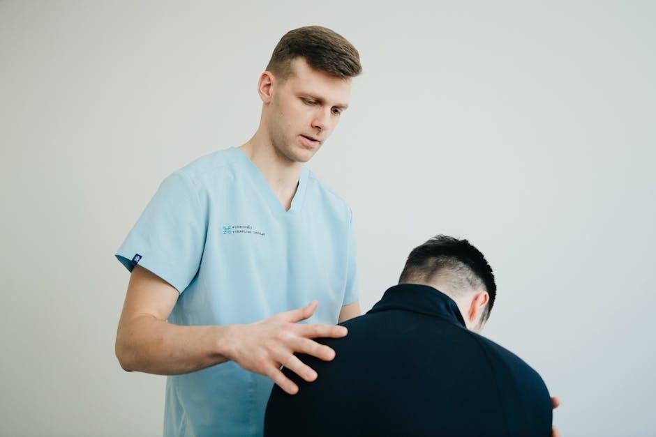
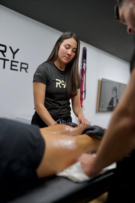
Pain Management & Edema Control
Effective pain management is paramount post-femur ORIF, enabling patient participation in rehabilitation. This typically involves a combination of pharmacological interventions, prescribed by the physician, and physical therapy modalities. Early implementation of pain control strategies is crucial for optimizing functional recovery.
Edema control is equally important, as swelling can impede range of motion and delay healing. RICE protocol – Rest, Ice, Compression, and Elevation – is frequently employed. Cryotherapy (ice packs) is applied regularly to reduce inflammation and pain.
Gentle range of motion exercises and soft tissue mobilization, performed by the physical therapist, also aid in edema reduction. Monitoring for signs of infection (fever, increased pain, redness) is essential, and any concerns should be immediately reported to the medical team.
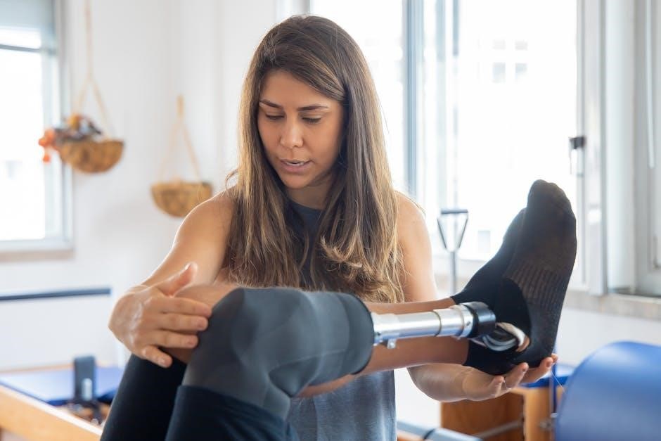
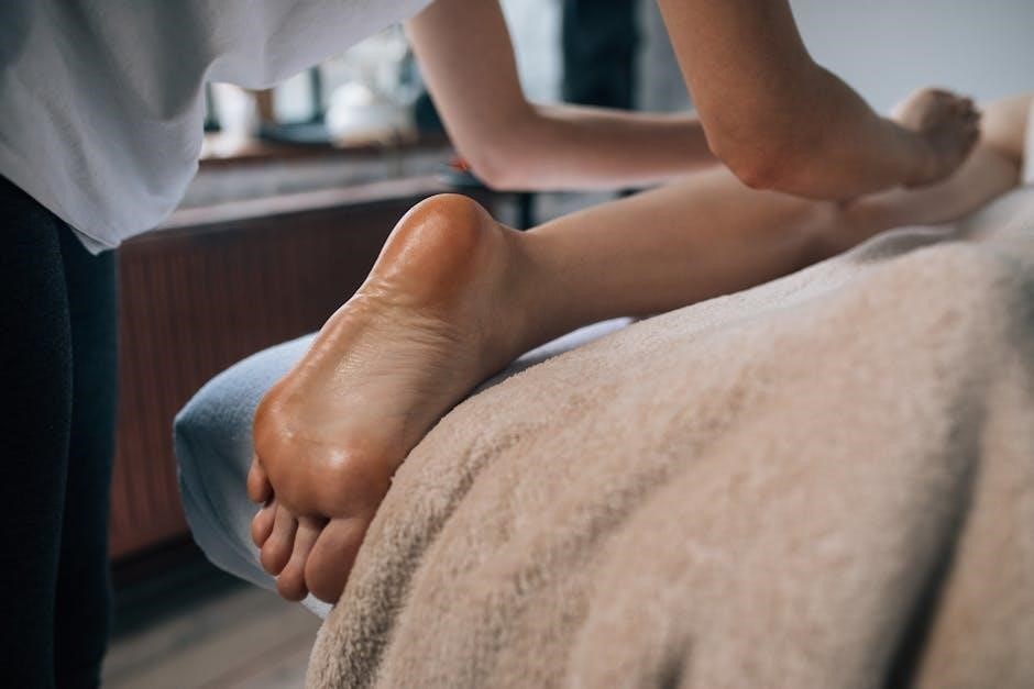
Early Range of Motion Exercises
Initiating gentle range of motion (ROM) exercises is vital in the immediate post-operative phase following femur ORIF, preventing stiffness and promoting joint mobility. These exercises are performed within the limitations set by the surgeon and immobilization protocol. Ankle pumps and gentle hip abduction/adduction are typically started immediately.
Heel slides and assisted knee flexion are introduced as pain allows, focusing on regaining knee extension and flexion. Emphasis is placed on pain-free movement, avoiding any forceful stretching or discomfort. The physical therapist will guide the patient through these exercises, ensuring proper form and technique.
Active-assisted range of motion, where the patient uses their own muscles with assistance from the therapist, is often utilized. Regular, consistent ROM exercises are crucial for maintaining joint integrity and preparing for more advanced rehabilitation phases.
Muscle Activation Exercises
Early muscle activation is paramount following femur ORIF surgery, focusing on restoring neuromuscular control without stressing the fracture site. Isometric quadriceps sets – tightening the thigh muscle without moving the knee – are initiated to maintain muscle mass and function. Gluteal squeezes are also crucial, activating the hip extensors and stabilizers.
Ankle pumps and gentle hip adduction/abduction further promote circulation and muscle engagement. Hamstring sets, gently pressing the heel into the bed, help activate the posterior thigh muscles. These exercises are performed frequently throughout the day, within a pain-free range.
The goal is to “wake up” the muscles surrounding the hip and knee, preparing them for more demanding strengthening exercises in subsequent phases. Proper technique and avoiding compensatory movements are emphasized by the physical therapist.
Phase 2: Early Rehabilitation (2-6 Weeks)
This phase emphasizes progressive weight-bearing, increased range of motion, and initial strengthening of the quadriceps, hip, and abductor muscles.
Gait training begins,
focusing on proper mechanics and minimizing limping.
Progressive Weight-Bearing
Weight-bearing progression is carefully managed during this phase (2-6 weeks post-ORIF). Initially, patients may continue with toe-touch weight-bearing, gradually advancing to partial weight-bearing (PWB) as tolerated, typically around weeks 2-4. This progression is guided by pain levels, fracture healing assessed via radiographs, and the patient’s ability to maintain proper biomechanics.
Physical therapists closely monitor for signs of instability or increased pain during weight-bearing activities. Assistive devices, such as crutches or a walker, are utilized to provide support and reduce stress on the healing fracture site. The goal is to achieve full weight-bearing (FWB) by week 6, but this timeline is individualized.
Emphasis is placed on proper gait mechanics to avoid compensatory patterns. Exercises to improve weight shifting and balance are incorporated. Patients are educated on weight-bearing precautions and instructed to avoid activities that could jeopardize fracture healing. Regular communication with the surgeon is essential to adjust the weight-bearing protocol as needed.
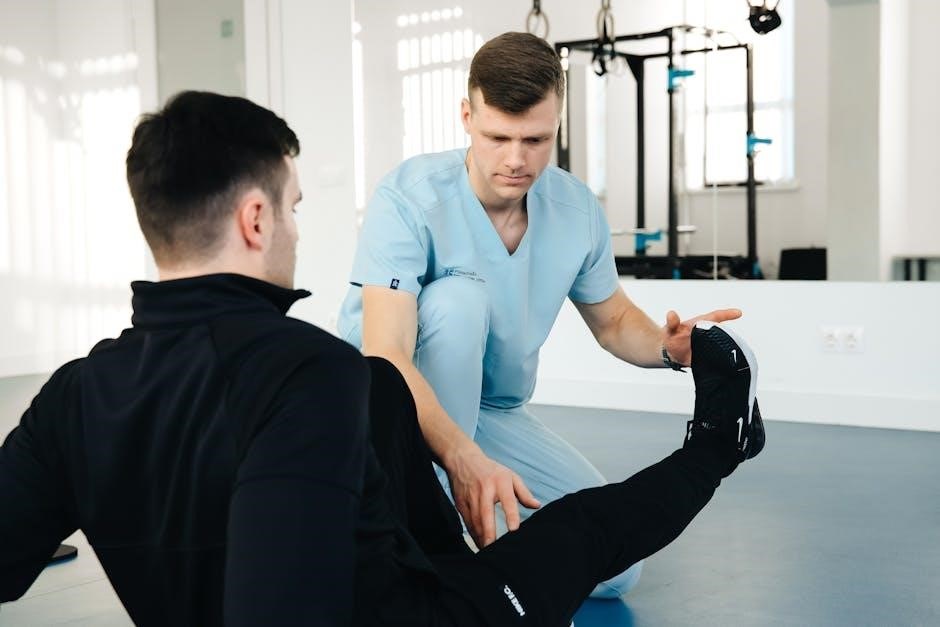
Range of Motion Progression
Restoring knee range of motion (ROM) is a priority during the early rehabilitation phase (2-6 weeks). Initially, gentle ROM exercises are performed within the limits of pain and immobilization, focusing on achieving full knee extension. Heel slides and assisted knee flexion are introduced to gradually increase flexion ROM.
As pain subsides and healing progresses, more aggressive ROM exercises are implemented. Stationary cycling without resistance can promote fluid mobilization and improve flexibility. Patellar mobilization techniques are utilized to address any restrictions. The goal is to achieve at least 90 degrees of knee flexion by week 6, with continued progression towards full ROM.
Physical therapists monitor for signs of inflammation or pain during ROM exercises and adjust the protocol accordingly. Patient education on home exercise programs is crucial to maintain ROM gains. Avoiding forceful stretching or overexertion is emphasized to prevent complications and ensure optimal healing.
Quadriceps Strengthening
Quadriceps strengthening is paramount for functional recovery following femur ORIF, beginning in Phase 2 (2-6 weeks). Initial exercises focus on isometric quadriceps contractions, performed in various knee angles, to activate the muscle without stressing the fracture site. These are held for 5-10 seconds, repeated multiple times throughout the day.
Progressing from isometrics, short-arc quadriceps exercises are introduced, utilizing ankle weights or resistance bands. Straight leg raises, performed in multiple planes, further enhance quadriceps strength and endurance. Emphasis is placed on VMO (vastus medialis obliquus) activation, crucial for patellar tracking and knee stability.
As strength improves, closed-chain exercises like mini-squats and leg presses are incorporated. Careful monitoring for pain and proper form is essential. The goal is to achieve at least 80% contralateral quadriceps strength before progressing to more advanced functional activities, ensuring a safe and effective rehabilitation process.
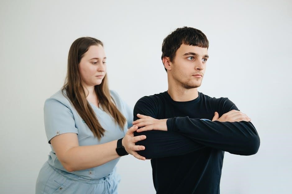
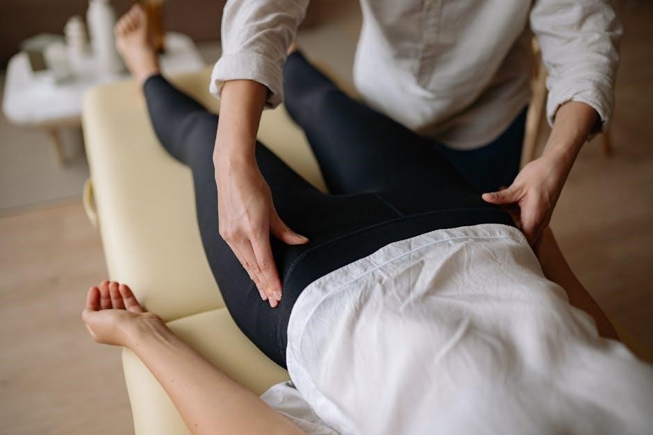
Hip & Abductor Strengthening
Hip and abductor strengthening is critical for restoring stability and proper gait mechanics post-femur ORIF, often initiated concurrently with quadriceps exercises. Initial focus involves isometric hip adduction, abduction, and extension, performed in pain-free ranges of motion. These exercises activate key stabilizing muscles without placing undue stress on the fracture.
Progressing from isometrics, resistance bands are utilized for hip abduction and external rotation exercises, targeting the gluteal muscles. Side-lying hip abduction, clam shells, and bridging exercises are incorporated to enhance abductor strength and endurance. Careful attention is given to maintaining proper form and avoiding compensatory movements.
As strength improves, single-leg stance exercises and step-ups are introduced, challenging balance and proprioception. Strengthening the hip abductors is vital for preventing Trendelenburg gait and ensuring a smooth, efficient walking pattern, contributing to overall functional recovery.
Gait Training – Initial Stages
Initial gait training focuses on establishing a safe and protected weight-bearing pattern following femur ORIF. Utilizing an assistive device – typically a walker or crutches – is paramount, adhering to the surgeon’s prescribed weight-bearing restrictions. Emphasis is placed on proper form, including heel-toe progression and symmetrical step length.
Early sessions involve short distances within a controlled environment, prioritizing quality over quantity. The physical therapist provides tactile cues and verbal guidance to correct deviations and promote efficient movement. Mirror practice can aid in visual feedback and motor learning.
Addressing gait abnormalities like limping or Trendelenburg gait is crucial. Exercises to improve hip and quadriceps strength are integrated into gait training. As tolerance increases, the distance and complexity of the gait pattern are gradually progressed, preparing the patient for more advanced stages.
Phase 3: Intermediate Rehabilitation (6-12 Weeks)
This phase builds strength and restores function, incorporating advanced exercises and proprioceptive drills.
Balance work and progressive gait training are key,
preparing for more demanding activities and a return to daily life.
Advanced Strengthening Exercises
During this phase (6-12 weeks), strengthening progresses beyond basic exercises to challenge the recovering leg. Focus shifts to functional movements mimicking daily activities. Exercises include closed-chain activities like squats (progressing depth as tolerated), lunges (initially short range), and step-ups onto varying heights.
Resistance is gradually increased using ankle weights, resistance bands, or weight machines. Specific attention is given to the vastus medialis obliquus (VMO), a crucial quadriceps muscle for knee stability. Hip abductor and adductor strengthening continues with side-lying exercises and resisted hip movements.
Core stabilization exercises are integrated to improve overall biomechanics and support the lower extremities. Plyometric exercises, such as controlled jumps and hops, may be introduced cautiously towards the end of this phase, assessing patient tolerance and stability. Regular monitoring of strength levels, aiming for 80% contralateral strength, guides progression.
Proprioceptive Training
Proprioception, or the body’s awareness of its position in space, is vital for regaining functional stability after femur ORIF. This phase (6-12 weeks) incorporates exercises designed to challenge and improve this sense. Single-leg stance activities are foundational, progressing from stable to unstable surfaces like foam pads or wobble boards.
Balance exercises include tandem stance, reaching activities in various directions, and perturbation training (controlled external disturbances). Neuromuscular electrical stimulation (NMES) can be used to facilitate muscle activation and enhance proprioceptive feedback.
Exercises involving dynamic movements, such as walking with head turns or catching a ball, further challenge the neuromuscular system. The goal is to restore the ability to react quickly and efficiently to changes in terrain or unexpected movements, reducing the risk of re-injury. Consistent practice and progression are key to optimizing proprioceptive function.
Balance Exercises
Balance deficits are common post-femur ORIF, impacting gait and functional independence. This phase (6-12 weeks) focuses on restoring static and dynamic balance. Initial exercises involve maintaining a stable base of support, progressing to narrower stances and eventually single-leg stance with minimal support.
Perturbation training – introducing controlled external forces – challenges the patient’s reactive balance control. Activities like reaching for objects while maintaining balance, and stepping in multiple directions, are incorporated. Use of balance aids (e.g., cane, walker) is gradually reduced as stability improves.
More advanced exercises include tandem stance, walking on uneven surfaces, and incorporating cognitive tasks during balance activities. The aim is to improve anticipatory and reactive balance strategies, enabling safe and efficient movement in various environments, ultimately reducing fall risk.
Gait Training – Advanced Stages
This phase (6-12 weeks) refines gait mechanics beyond basic ambulation, focusing on efficiency and normalization. Emphasis shifts to improving stride length, cadence, and symmetry. Hill work and stair climbing are introduced gradually, challenging strength and endurance.
Proprioceptive feedback is crucial; exercises incorporating varied surfaces (grass, gravel) enhance awareness and control. Functional gait training simulates real-life activities like navigating obstacles and carrying objects. Video gait analysis can identify remaining asymmetries and guide corrective strategies.
The goal is to achieve a near-normal gait pattern with minimal compensatory movements. Patients progress to more demanding activities like jogging or running (if appropriate), preparing for a return to higher-level function. Regular monitoring ensures proper form and prevents re-injury.
Phase 4: Late Rehabilitation & Return to Function (12+ Weeks)
This final stage prioritizes functional exercise, simulating activities specific to the patient’s goals. A gradual return to activity is guided by strength, range of motion, and pain levels.
Long-term maintenance programs prevent re-injury and optimize performance, addressing potential complications like stiffness or weakness.
Functional Exercises
Functional exercises bridge the gap between controlled rehabilitation and real-world activities, focusing on movements required for daily living and desired recreational pursuits. These exercises are progressively challenging, mimicking tasks like stair climbing, squatting, lunging, and walking on uneven surfaces.
Initially, these activities are performed with close supervision, emphasizing proper form and biomechanics to prevent compensatory patterns. Progression involves increasing repetitions, adding resistance (using weights, resistance bands, or body weight), and decreasing the level of assistance.
Specific exercises may include step-ups, lateral step-downs, balance activities on unstable surfaces, and agility drills. The goal is to restore neuromuscular control, improve dynamic stability, and enhance the patient’s confidence in performing functional tasks independently. A home exercise program is crucial for maintaining gains and promoting long-term functional improvements.
Careful monitoring for pain, swelling, or fatigue is essential, and exercises should be modified accordingly.
Return to Activity Progression
Returning to activity after femur ORIF is a gradual process guided by established criteria and individual patient factors. Initial stages focus on low-impact activities like walking on level surfaces and stationary cycling, gradually increasing duration and intensity. Progression to higher-impact activities, such as jogging, running, and jumping, requires adequate strength, range of motion, and neuromuscular control.
A structured progression is essential, typically involving a phased approach with specific milestones to achieve before advancing. Criteria for advancement may include achieving a certain percentage of contralateral strength, demonstrating pain-free range of motion, and successfully completing functional exercises.
Sport-specific training is incorporated as appropriate, focusing on movements and demands relevant to the patient’s desired activities. Close monitoring for signs of pain, swelling, or fatigue is crucial, and activity levels should be adjusted accordingly. Patient education regarding proper warm-up, cool-down, and injury prevention strategies is paramount.
Realistic expectations and adherence to the rehabilitation program are key to a successful return to activity.
Long-Term Maintenance
Long-term maintenance following femur ORIF is crucial for sustained functional recovery and preventing re-injury. Continued adherence to a home exercise program, focusing on strength, flexibility, and proprioception, is essential. Regular cardiovascular exercise, such as walking, swimming, or cycling, helps maintain overall fitness and endurance.
Periodic reassessment by a physical therapist can identify any deficits or imbalances that may develop over time. Activity modification may be necessary to accommodate changes in lifestyle or activity demands. Maintaining a healthy weight reduces stress on the operated limb and promotes joint health.
Patient education regarding proper body mechanics and injury prevention strategies remains vital. Listening to the body and avoiding overexertion are key to preventing flare-ups or complications. Proactive management of any pain or discomfort is essential for long-term success.
Consistent effort in maintaining these principles will optimize functional outcomes and quality of life.
Potential Complications & Red Flags
Post-femur ORIF, several complications require prompt attention. Infection, indicated by fever (over 101°F, though low-grade is normal initially), increased pain, redness, or drainage from the incision, demands immediate medical evaluation. Deep vein thrombosis (DVT), presenting as calf pain, swelling, or warmth, necessitates urgent assessment.
Non-union or malunion of the fracture, evidenced by persistent pain or instability, may require further surgical intervention. Hardware failure, such as screw loosening or plate breakage, can cause pain and functional limitations. Nerve or vascular injury, manifesting as numbness, tingling, or changes in circulation, requires immediate attention.
Red flags during rehabilitation include sudden increases in pain, swelling, or stiffness, as well as any signs of instability. Report any concerns to your physical therapist or surgeon promptly. Early identification and management of complications are crucial for optimal outcomes.